Rv Lv Ratio Measurement Ct

Ctpa Demonstrating The Rv Lv Ratio Measurement Note Ctpa Computed Download Scientific Diagram

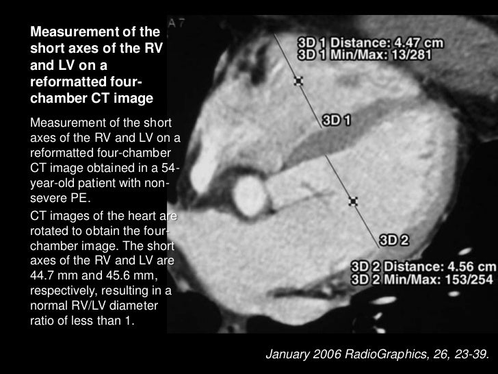
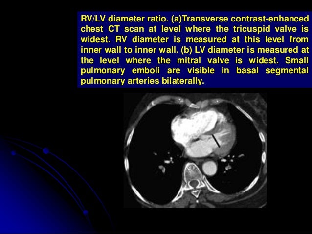
Measurement Of Rv Lv Ratio Axial Ct Images Demonstrate The Best Download Scientific Diagram

Amgrad Rv Lv Ratio
Http Pdf Posterng Netkey At Download Index Php Module Get Pdf By Id Poster Id 127655

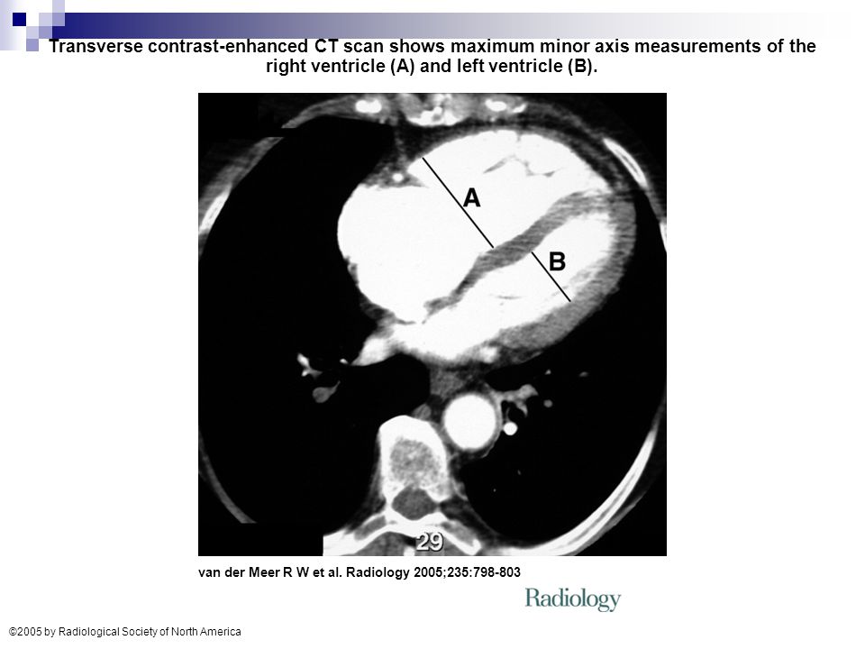
Transverse Contrast Enhanced Ct Scan Showing Maximum Minor Axis Download Scientific Diagram

Moderate Acute Dilatation Of The Rv In A 55 Year Old Man With Massive Download Scientific Diagram
Pvr 2 8 1 8 3 1 wood.
Rv lv ratio measurement ct.

Rv Lv Diameter Ratio 1 Not Associated With Worse Outcomes In Acute Pe Pulmonology Advisor

Transverse Contrast Enhanced Ct Scan Shows Maximum Minor Axis Download Scientific Diagram
Http Pdf Posterng Netkey At Download Index Php Module Get Pdf By Id Poster Id 120662

Amgrad Chamber Sizes On Chest Ct
Management Of Massive Submassive Pulmonary Embolism

What S New In Emergency Radiology At Harborview Uw Emergency Radiology
Https Journal Chestnet Org Article S0012 3692 19 31374 1 Pdf
Imaging Of Pulmonary Embolism

Ct Angiogram Of The Heart At The Level Of The Midventricles Shows Download Scientific Diagram
Acute Pulmonary Embolism Diagnosis And Management Ppt Video Online Download

Repeated And Adaptive Multidisciplinary Assessment Of A Patient With Acute Pulmonary Embolism And Recurrent Cardiac Arrests Bmj Case Reports

Right Ventricular To Left Ventricular Ratio At Ct Pulmonary Angiogram Predicts Mortality In Interstitial Lung Disease Chest

Pulmonary Hypertension Radiology Reference Article Radiopaedia Org

Imaging Right Left Ventricular Interactions Jacc Cardiovascular Imaging

Is Ct Angiography Derived Right To Left Ventricular Diameter Ratio Adequate For Pulmonary Embolism Risk Stratification Jacc Journal Of The American College Of Cardiology

Measurement Of Right And Left Ventricular Diameter Download Scientific Diagram
Pulmonary Hypertension Radiology Key
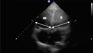
Echocardiographic Findings Of Pulmonary Embolism In The Parasternal Download Scientific Diagram

Reproducibility Of Ct Signs Of Right Ventricular Dysfunction In Acute Pulmonary Embolism Semantic Scholar

Rvlv Imbio

5 Pulmonary Embolism Radiology Key
Submassive Pe Emory School Of Medicine

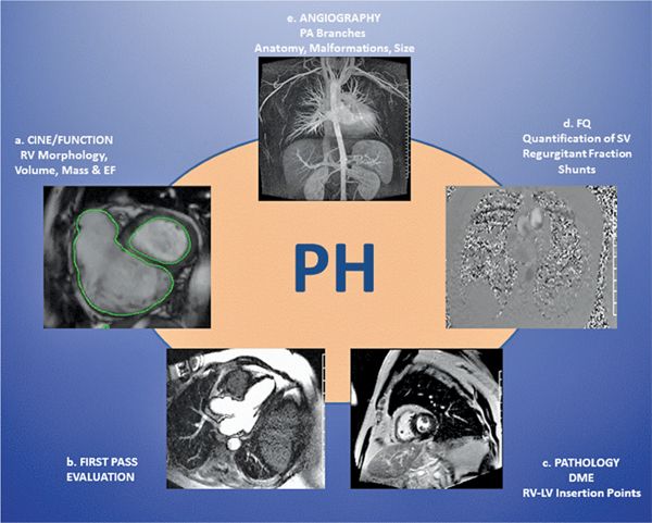
Findings Of Cardiac Magnetic Resonance Imaging Download Table

Imaging Right Left Ventricular Interactions Sciencedirect
Source : pinterest.com
